Victorian Anatomical Atlases and Their Many Lives (and Deaths)
Victorian Anatomical Atlases and Their Many Lives (and Deaths)
By Keren Rosa Hammerschlag
Introducing Joseph Maclise, Victorian Anatomist
It was 2007, and I was a PhD student sitting in the special collections room in the Wellcome Library, London, searching for nineteenth-century British images of dissections. Victorian artists and art students most certainly attended, and sometimes even participated in, dissections, but I was struggling to find any visual evidence of it. It was fast becoming apparent that human dissection was considered an inappropriate subject for artistic treatment, and a rather unacceptable undertaking for any respectable Victorian gentleman.1 Noticing my frustration, William Schupbach, lead librarian and curator at the Wellcome, presented me with the first British, 1851, folio edition of Joseph Maclise’s Surgical Anatomy (fig. 1). This little-known anatomical production is the jumping-off point for the current One Object feature, “Victorian Anatomical Atlases and Their Many Lives (and Deaths)”.
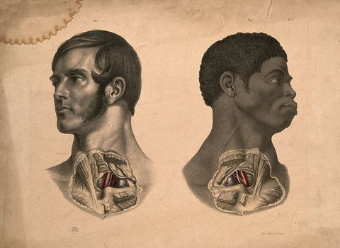
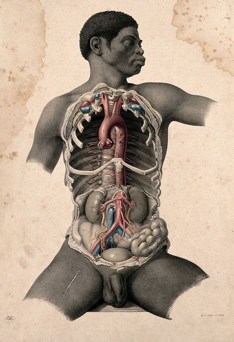
1One particularly significant aspect of Maclise’s Surgical Anatomy is that it includes two illustrations of the dissection of a Black man: Plates 5 and 14 of the first British edition (figs. 2 and 3). I examine Plate 14 in depth in my article for this feature, “Black Apollo: Aesthetics, Dissection, and Race in Maclise’s Surgical Anatomy”. Years after first encountering Surgical Anatomy in the Wellcome Library, I was in the National Library of Medicine in Bethesda, Maryland, USA, looking for the dissected Black man, only to discover that he was missing from all American editions of the same atlas (figs. 4, 5, and 6). The discovery of this mysterious transatlantic erasure was developed into a paper, which I delivered in May 2018 at the “Objects in Motion” workshop in Giverny, France, sponsored by the Terra Foundation for American Art, the Paul Mellon Centre for Studies in British Art, and the Yale Center for British Art. It soon became apparent that a comprehensive analysis of the British and American editions of Maclise’s Surgical Anatomy required more voices than just mine. With support from the Terra Foundation, along with Angela McShane, Julia Nurse, and William Schupbach at the Wellcome, and Baillie Card and Sarah Victoria Turner at the Paul Mellon Centre, in April 2019, a remarkable group of historians, art historians, medical historians, curators, and librarians gathered in London to examine “in the flesh” the Wellcome Library’s impressive collection of eighteenth- and nineteenth-century anatomical atlases. The ideas generated by that event formed the basis of the current feature, and I am deeply grateful to everyone who believed in the project and worked to bring it to fruition, above all, the editors of British Art Studies.
Joseph Maclise was born in Cork, Ireland, in 1815. In the 1830s, he studied at University College, London, under Robert Liston and Samuel Cooper, to whom he dedicated Surgical Anatomy. He also studied in Paris at the École Pratique, L’Hôpital de la Pitié. His brother was the successful history painter, Daniel Maclise, and the two travelled together to Paris and other European cities in 1844. Interestingly, several sets of brothers make appearances over the course of this feature: the Maclise brothers, of course, Charles and John Bell, Richard and Jones Quain, and William and John Hunter. Before setting out for Europe with his brother, Maclise produced the illustrations for his former teacher, Richard Quain’s The Anatomy of the Arteries of the Human Body (1844).2 In Comparative Osteology of 1847, which Maclise wrote and illustrated, he identified what he termed the “archetype” skeleton, or the complete form from which different skeletal structures derived.3 He claimed to have come up with the concept—or at least this particular use of the term “archetype”—prior to the influential comparative anatomist, and opponent of Charles Darwin’s theory of evolution by natural selection, Richard Owen.4 Nonetheless, Maclise’s contribution to the study of human and animal anatomy continues to languish in obscurity. Following the publication of the second American edition of Surgical Anatomy in 1859, he published On Dislocations and Fractures and, by the 1860s, had fallen afoul of the medical fraternity.5 At this point, the archival trail runs cold, except for Michael Sappol’s remarkable discovery of Maclise’s death certificate (fig. 7). No obituaries have yet been found.
2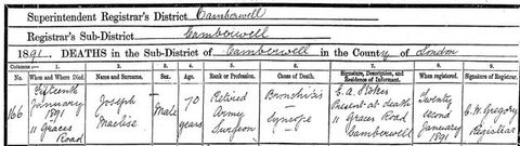
Maclise’s Surgical Anatomy was initially released in four parts, starting in 1848, as an imperial folio with individual fasciculus. In 1851, John Churchill of London, and Blanchard and Lea of Philadelphia, published the atlas as complete first editions. In 1856, Churchill published a second expanded edition and, in 1859, Blanchard and Lea did the same. Henry C. Lea published a smaller, cheaper version in 1870. The atlas presents a series of illustrations of dissections with the purpose of teaching surgeons and aspiring surgeons the anatomical structures relevant to the successful performance of surgical procedures. The preface begins as follows: “The object of this work is to present to the student of medicine and the practitioner removed from the schools, a series of dissections demonstrative of the relative anatomy of the principal regions of the human body.”6 The surface of the human body, Maclise argued, was like a map; the surgeon was required to read the topography of the body, seeing through the skin to the anatomy beneath. He called on his surgical readers to assume an “expansive gaze” and engage in a form of comparative anatomy—viewing human anatomy in relation to “all allying and allied species”.7 “Comparison may be fairly termed the pioneer to all certain knowledge,” he wrote.8
6The monumental undertaking of producing the images and text for Surgical Anatomy was likely intended to establish Maclise as an eminent anatomist and anatomical illustrator in his own right. Nonetheless, it is tempting to imagine his Royal Academician brother, Daniel, assisting with this large artistic undertaking. The illustrations in Surgical Anatomy certainly work to demonstrate the artistic skill of their maker(s), featuring depictions of ideal physical specimens: attractive, seemingly healthy men, in their prime. Aside from their developed musculature, they bear no evidence of hard labour, poverty, or illness—they look more asleep than dead. On occasion, their poses are even made to invoke classical statues such as the Belvedere Torso, thereby elevating the atlas to the status of “high” art.
Despite its artistic and scientific merits, Maclise’s Surgical Anatomy has been overlooked largely in both the art-historical and medical-historical literature.9 At best, it is briefly mentioned in histories of anatomical illustration.10 One possible explanation for this oversight is that Maclise’s atlas is easily dismissed as too medical for art historians and too artistic for medical historians. Additionally, it does not fit seamlessly into traditional narratives about the development of anatomical illustration. After all, Henry Gray’s Anatomy of the Human Body, with its modern-looking, pared down illustrations, was first published in 1858—only seven years after the publication of the complete first edition of Surgical Anatomy. It is difficult to square Maclise’s large, elaborate, colourful engravings with Henry Vandyke Carter’s starker productions. Therefore, it is not surprising that Surgical Anatomy received a lukewarm reception in the United Kingdom when it was first published. The moment for these kinds of anatomical productions in the British context, it seems, had passed.
9We often talk of canonical artists as being ahead or of their time; with Maclise, it feels as though we are dealing with a great talent who came a moment too late. Hence, in an effort to raise Maclise and his work from obscurity, this One Object feature brings together historians of art and historians of medicine in what has proven to be an exciting exercise in interdisciplinarity. A series of three short films featuring Ludmilla Jordanova and William Schupbach in conversation in the Wellcome Library focus on Maclise’s productions, the contexts in which they were made, used, and collected, and the materiality of the objects themselves. In his article, “Joseph Maclise, Taylor & Walton, and Publishing on Gower Street in the 1840s”, Schupbach maps medical publishing and publishers around Gower Street in Bloomsbury during the nineteenth century. Anthea Callen, in “Bloodlines: Circulating the Male Body Across Borders in Art and Anatomy 1780–1860”, situates Maclise in the medical and artistic networks of nineteenth-century Ireland, England, and France, in order to demonstrate the impact on Maclise’s illustrations of a variety of visual sources. Naomi Slipp reveals in her article, “‘It Should Be On Every Surgeon’s Table’: The Reception and Adoption of Joseph Maclise’s Surgical Anatomy (1851) in the United States”, that Maclise’s Surgical Anatomy was remarkably well received in the United States. Michael Sappol’s richly illustrated article, “Mr Joseph Maclise and the Epistemology of the Anatomical Closet”, offers a provocative examination of the “queerness” of Maclise’s illustrations, character, and relationships, and the palpable homoeroticism of several of his illustrations.
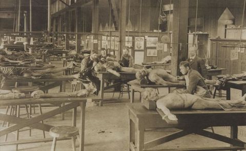
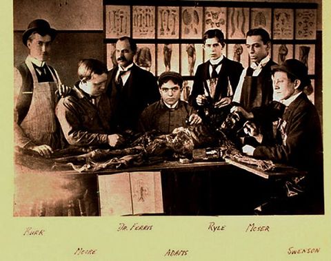
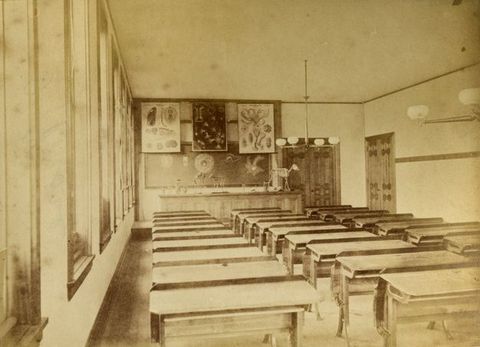
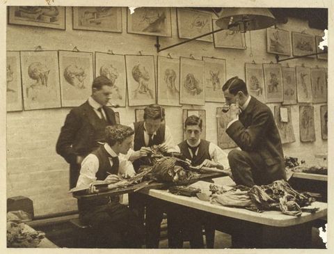
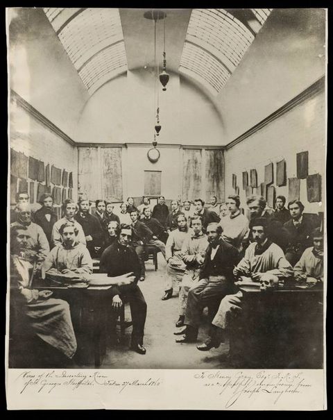
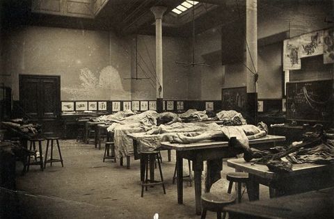
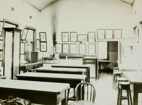
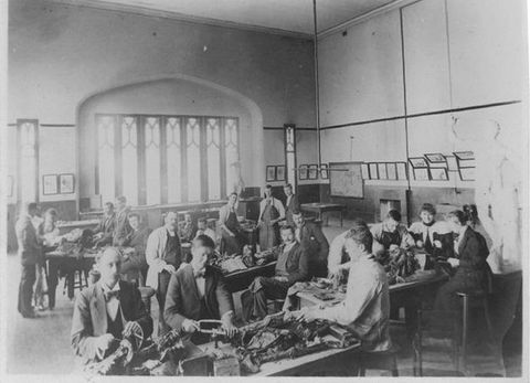
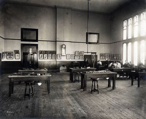
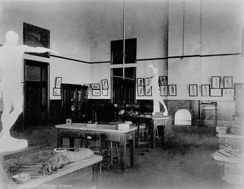
Despite the sanitised nature of many of Maclise’s illustrations, it is clear that we are looking at dead bodies. Hence, I encourage readers to proceed with care. The substantial number of photographs that were taken of anatomy theatres and dissecting rooms during the nineteenth century offer vital clues to the use of illustrations of dissections, such as those produced by Maclise.11 These photographs, which can be difficult to look at, invariably feature objects: skeletons, écorchés, casts of classical statues and statuettes, and illustrations of dissections hanging on the walls; they also sometimes include people—dead and alive. While far from comprehensive, photographs of British, American, and Australian anatomy theatres are included here in an image gallery. Some of these photographs are discussed by contributors to this One Object feature; others are included to give a sense of the environments in which dissections were performed and the kinds of objects that were used for anatomical instruction during the nineteenth and early twentieth century (figs. 8–18). Looking at these photographs, it is essential that we remain mindful of the ethical concerns surrounding the public and private display of images and sculptures of dead and anatomised bodies. As several of the articles in this feature make clear, histories of anatomical illustration and modelling are inevitably bound up with the fraught issues of consent, exploitation, voyeurism, and the status of the corpse.
11Finally, gaps remain for future scholars to fill in. Much is still unknown about Maclise. The nature of his relationship with his brother Daniel is commented on by all of the contributors, but the biographical material is one sided; Daniel’s life has been written, Joseph’s has not.12 As a result, we are often forced to find Joseph in Daniel’s biography. Above all, with very little to work with aside from the images and texts produced by (white, male) doctors and artists like Maclise, we continue—slowly—to piece together the identities of the men, women, and children, who ended up on the dissecting table against their will in an age before consent was required to cut open dead bodies for the purpose of “Anatomical Examination”.13
12
Acknowledgements
This project is made possible through support from the Terra Foundation for American Art.
About the author
-
 Keren Rosa Hammerschlag is a lecturer in art history and curatorship in the Centre for Art History and Art Theory, School of Art and Design, at the Australian National University, Canberra. Her current research focuses on nineteenth-century anatomical illustration, and race in Victorian painting. She is the author of Frederic Leighton: Death, Mortality, Resurrection (Aldershot: Ashgate, 2015) and “Christ’s Racial Origins: Finding the Jewish Race in Victorian History Painting”, an article published in The Art Bulletin in 2021.
Footnotes
-
1
I elaborate on this point in Keren Rosa Hammerschlag, “The Gentleman Artist-Surgeon in Late Victorian Group Portraiture”, Visual Culture in Britain 14, no. 2 (2013): 154–178. ↩︎
-
2
Richard Quain, The Anatomy of the Arteries of the Human Body with its Applications to Pathology and Operative Surgery in Lithographic Drawings with Practical Commentaries: The Drawings from Nature and on Stone by Joseph Maclise (London: Taylor and Walton, 1844). Quain writes in the Preface: “To carry out my views as to the delineations, I obtained the assistance of my friend and former pupil, Mr. Joseph Maclise”. ↩︎
-
3
Joseph Maclise, Comparative Osteology: Being Morphological Studies to Demonstrate the Archetype Skeleton of Vertebrated Animals (London: Taylor and Walton, 1847). I examine this text in greater depth in Keren Rosa Hammerschlag, “Drawing Racial Comparisons in Nineteenth-Century British and American Anatomical Atlases”, in Victorian Science and Imagery: The Evolution of Form in Nineteenth-Century Visual Culture (Pittsburgh, PA: University of Pittsburgh Press, forthcoming 2021). See also Adrian Desmond, The Politics of Evolution: Morphology, Medicine, and Reform in Radical London (Chicago, IL: University of Chicago Press, 1989). ↩︎
-
4
Nicolaas A. Rupke, “Richard Owen’s Vertebrate Archetype”, Isis 84, no. 2 (1993): 234–235. ↩︎
-
5
Joseph Maclise, On Dislocations and Fractures (London: John Churchill, 1859). As Michael Sappol outlines in his contribution to this One Object feature, Maclise inserted into On Dislocations and Fractures an “off-kilter diatribe against William Harvey’s account of the action of the heart”. Maclise then attempted, unsuccessfully, to defend his position in letters sent to the Lancet. After this, his name falls out of the record. [We will add Mike’s reference when ready]. ↩︎
-
6
Joseph Maclise, Preface to Surgical Anatomy (Philadelphia, PA: Blanchard and Lea, 1851), v. ↩︎
-
7
Maclise, Preface to Surgical Anatomy, v. ↩︎
-
8
Maclise, Preface to Surgical Anatomy, vi. I elaborate on these ideas in “Drawing Racial Comparisons in Nineteenth-Century British and American Anatomical Atlases”. ↩︎
-
9
Two exceptions are Rebecca E. May, “‘This Shattered Prison’: Bodily Dissolution, Wuthering Heights, and Joseph Maclise’s Dissection Manuals”, Nineteenth-Century Contexts 33, no. 5 (2011): 415–436; and The Anatomy Lesson: Art and Medicine, An Exhibition of Art and Anatomy to Celebrate the Tercentenary of the Royal Charter of 1692 of the Royal College of Physicians of Ireland (Dublin: The National Gallery of Ireland, 1992), 31–32 and 74–76. ↩︎
-
10
Illustrations from Surgical Anatomy are discussed in K.B. Roberts and J.D.W. Tomlinson, The Fabric of the Body: European Traditions of Anatomical Illustration (Oxford: Clarendon, 1992), 570–573. ↩︎
-
11
Historian of medicine, John Harley Warner, analyses photographs of American medical students gathered around cadavers in Dissection: Photographs of a Rite of Passage in American Medicine 1880–1930, ed. J.H. Warner and James M. Edmonson (New York: Blast Books, 2009). ↩︎
-
12
William Justin O’Driscoll, A Memoir of Daniel Maclise, R.A. (London: Longmans, Green, and Co., 1871); Richard Ormond, “Daniel Maclise”, Burlington Magazine 110 (December 1968): 684–693; Nancy Weston, Daniel Maclise: An Irish Artist in Victorian London (Dublin: Four Courts Press, 2001); and Peter Murray, ed., Daniel Maclise 1806–1870: Romancing the Past, exhibition catalogue (Cork: Crawford Art Gallery, 2008). ↩︎
-
13
The 1832 British Anatomy Act made unclaimed human bodies legally available to medical schools for “Anatomical Examination”. An Act for Regulating Schools of Anatomy [1st August 1832]. The National Archive, UK. https://www.nationalarchives.gov.uk/education/resources/body-snatchers/source-four-the-anatomy-act-1832/. ↩︎
Imprint
| Author | |
|---|---|
| Date | 18 July 2021 |
| Category | One Object |
| Review status | Peer Reviewed (Double Blind) |
| License | Creative Commons Attribution-NonCommercial 4.0 International (CC BY-NC 4.0) |
| Downloads | PDF format |
| Article DOI | https://doi.org/10.17658/issn.2058-5462/issue-20/1objintro |
| Cite as | Hammerschlag, Keren Rosa. “Victorian Anatomical Atlases and Their Many Lives (and Deaths).” In British Art Studies. London and New Haven: Paul Mellon Centre for Studies in British Art and Yale Center for British Art, 2021. https://doi.org/10.17658/issn.2058-5462/issue-20/1objintro. |